
Figure 6
- ID
- ZDB-FIG-191230-1578
- Publication
- Johansson et al., 2019 - Dkk1 Controls Cell-Cell Interaction through Regulation of Non-nuclear β-Catenin Pools
- Other Figures
- All Figure Page
- Back to All Figure Page
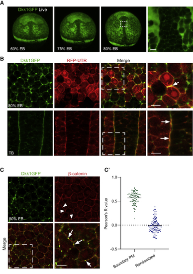
Ubiquitous Expression of GFP-Tagged Dkk1 Reveals Polarized Distribution of Dkk1 Protein at Tissue and Subcellular Levels (A) Live (B) Subcellular localization of Dkk1GFP in 80% epiboly (EB) and tail bud (TB) stage fixed embryos. Single-plane high magnification confocal images are shown of the region of interest in dashed white frames. Localization of Dkk1GFP to actin-rich membrane junctions was visualized by coexpression with red fluorescent protein-tagged actin-binding utrophin (RFP-UTR, red). Arrow highlights Dkk1GFP and RFP-UTR colocalization at a cell-cell junction. n = 6 for each condition. Scale bar, 20 μm. (C) Dkk1GFP colocalizes with plasma membrane-associated β-catenin (red)-positive puncta. Embryos were fixed shortly after heat shock. Arrowheads highlight β-catenin positive puncta (red), which colocalize with Dkk1GFP (arrows). (C’) Quantification of colocalization (n = 10). Mean Pearson’s R value is 0.60 for Dkk1-β-catenin and 0.03 for the randomized control. Scale bar, 20 μm. |
| Gene: | |
|---|---|
| Antibody: | |
| Fish: | |
| Condition: | |
| Anatomical Terms: | |
| Stage Range: | Shield to Bud |
Reprinted from Developmental Cell, 51(6), Johansson, M., Giger, F.A., Fielding, T., Houart, C., Dkk1 Controls Cell-Cell Interaction through Regulation of Non-nuclear β-Catenin Pools, 775-786.e3, Copyright (2019) with permission from Elsevier. Full text @ Dev. Cell