
Fig. S2
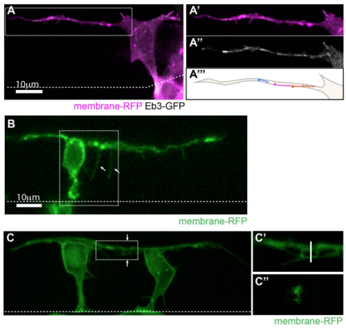
Basal protrusions contain dynamic microtubules, form filopodia and can overlap (related to Figure 1). A) T-shaped differentiating neuron with a long basal protrusion. White box marks the area of higher magnification shown in A’-A’’’). A’) Membrane-RFP. A’’) EB3-GFP (Movie 2). A’’’) Illustration of EB3-GFP comet trajectories during a 30 second timelapse period. B) T-shaped cell with filopodia (arrows) on basal protrusion (Movie 3). C) Two neurons differentiating 18µm apart with overlapping basal arms. C’) Higher magnification of white box in C). C’’) Cross-section of the basal protrusions at the line shown in C’). All images are projected images from confocal z-stacks. Dashed line shows the apical surface. |
Reprinted from Developmental Cell, 49, Hadjivasiliou, Z., Moore, R.E., McIntosh, R., Galea, G.L., Clarke, J.D.W., Alexandre, P., Basal Protrusions Mediate Spatiotemporal Patterns of Spinal Neuron Differentiation, 907-919.e10, Copyright (2019) with permission from Elsevier. Full text @ Dev. Cell