Fig. S2
- ID
- ZDB-FIG-190716-6
- Publication
- Han et al., 2019 - Vitamin D Stimulates Cardiomyocyte Proliferation and Controls Organ Size and Regeneration in Zebrafish
- Other Figures
- All Figure Page
- Back to All Figure Page
|
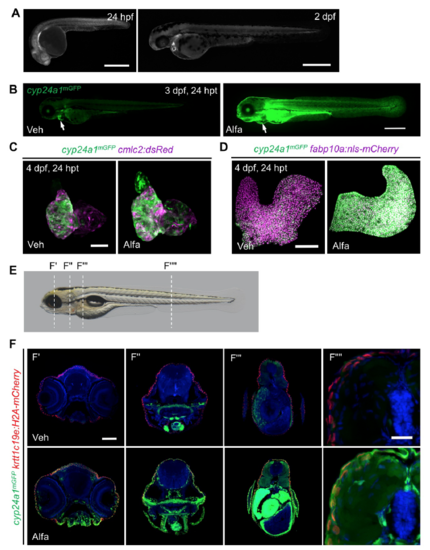
cyp24a1mGFP Reporter Fish Indicate Vitamin D Activity (Related to Figure 2) (A) mGFP fluorescence in 24 hpf and 48 hpf cyp24a1mGFP embryos. Scale bar, 500 μm. (B) Lateral view of a cyp24a1mGFP reporter knock-in at 3 dpf following treatment with vehicle or Alfa for 24 hours. Arrowheads indicate cardiac mGFP fluorescence. Scale bar, 500 μm. (C) Maximum intensity projection images of dissected 4 dpf cmlc2:dsRed;cyp24a1mGFP hearts after vehicle or Alfa treatment for 24 hours. Scale bar, 50 μm. (D) Maximum intensity projection images of dissected 4 dpf fabp10a:nls-mCherry; cyp24a1mGFP livers (to mark hepatocytes) after vehicle or Alfa treatment for 24 hours. Scale bar, 100 μm. (E) Representative 4 dpf wild-type embryo from Figure 2B indicating section plane of Figures F’ to F’’’’ with dashed lines. (F) Maximum projections of 10 μm confocal sections at indicated planes of 4 dpf krtt1c19e:H2A-mCherry; cyp24a1mGFP (to mark basal epithelial cells) embryos treated with vehicle or Alfa. Scale bars, 100 μm for F’-F’’’ and 20 μm for F’’’’. |
Reprinted from Developmental Cell, 48(6), Han, Y., Chen, A., Umansky, K.B., Oonk, K.A., Choi, W.Y., Dickson, A.L., Ou, J., Cigliola, V., Yifa, O., Cao, J., Tornini, V.A., Cox, B.D., Tzahor, E., Poss, K.D., Vitamin D Stimulates Cardiomyocyte Proliferation and Controls Organ Size and Regeneration in Zebrafish, 853-863.e5, Copyright (2019) with permission from Elsevier. Full text @ Dev. Cell

