
Fig. 3
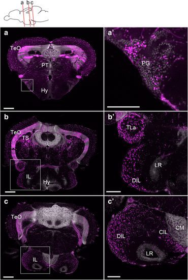
Localization of the mCherry-positive cells in the adult brain of Tg(her5:ERT2CreERT2;βact:lox-stop-lox-hmgb1:mCherry) zebrafish treated with tamoxifen at 24 hpf. a–c Confocal images of frontal sections showing global views of the mCherry distribution (Z-projection, 5 μm for a and 10 μm for b and c). The mCherry-positive cells are shown in magenta, and DAPI nuclear labeling is shown in gray. The plane of each section is indicated in the schematic drawing on the top. a’–c’ Higher magnifications of the areas squared in a–c, showing the preglomecular nucleus (PG; a’) and the inferior lobe (IL; b’, c’). Abbreviations: CM corpus mamillare, CIL central nucleus of the inferior lobe, DIL diffuse nucleus of the inferior lobe, Hy hypothalamus, IL inferior lobe, LR lateral recess, PG preglomerular nucleus, PT pretectum, TeO optic tectum, TLa torus lateralis, TS torus semicircularis. Scale bars: a–c, 200 μm; a’–c’, 100 μm |