
Fig. 9
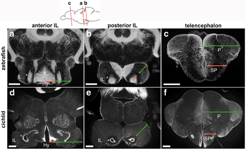
Comparison of the zebrafish and cichlid brains. Frontal sections of the brains of zebrafish (a–c) and cichlid (d–f), showing DAPI nuclear labeling in gray. The plane of the zebrafish sections is indicated in the schematic drawing on the top, and comparable level of the cichlid brain is shown below each zebrafish section. a, d Anterior IL. b, e The posterior IL. c, f The telencephalon. The relative size of the cichlid IL (d, e) is much larger than that of the zebrafish IL (a, b). It is prominent in comparison with the size of the hypothalamus (Hy; the size indicated in red arrows in a and d) that is located medial to the IL (the size indicated in green arrows in a and d). Also, the relative size of the external zone (the size indicated in green arrows in b and e) in comparison with the internal ventricular zone (the size indicated in red arrows in b and e) is much larger in cichlid. The asterisks (*) in a, b, d, and e indicate a cell-free fiber zone separating the external and internal zones. The relative size of the pallium (P; the size indicated in green arrows in c and f) in comparison with the subpallium (SP; the size indicated in red arrows in c and f) is much larger in cichlid than in zebrafish. Scale bar: a-c, 200 μm; d–f, 350 μm. Abbreviations: Hy hypothalamus, IL inferior lobe, P pallium, SP subpallium |