
Fig. 6
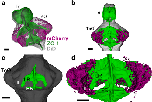
Developing IL in relation to the ventricular morphology. 3D reconstruction of image segmentation from confocal images of a 14 dpf zebrafish brain. ZO-1 (ventricular labeling) is shown in green (a–d), mCherry-positive cells are shown in magenta (a, b, d), and DiD fiber labeling is shown in gray (a–c). a, b Oblique (a) and ventral (b) views of the brain, showing the general distribution of the mCherry-positive cells in relation to the ventricular organization. c A ventral view highlighting the lateral recess (LR) and the posterior recess (PR) (anterior of the brain to the top). d A higher magnification of bfocusing on the mCherry cells in relation to the LR. The mCherry cells are continuous from the tectal region, but they are devoid of proximity of the ventricular zone. Scale bars, 50 μm. Abbreviations: IL inferior lobe, LR lateral recess, PR posterior recess, Tel telencephalon, TeO optic tectum |