
Fig. 10
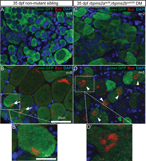
Bucky ball stained structures are abnormal in rbpms2 mutants. (A) Non-mutant sibling males have no specific Buc signal. (B) Non-mutant sibling female with asymmetric Buc localization in compact perinuclear domain of primary (stage I) oocytes (arrows). (B?) Magnified view of normal Buc localization in non-mutant sibling oocyte. (C) Like their control male siblings, differentiated male rbpms2 mutants have no Buc signal. (D) Oocyte-like cells of rbpms2 mutant gonads have more dispersed asymmetric Buc staining, frequently observed in a ring-like pattern (arrow-heads). (D?) Magnified view of ring-like Buc localization in rbpms2 mutant oocyte. |
| Gene: | |
|---|---|
| Fish: | |
| Anatomical Term: | |
| Stage: | Days 30-44 |