
FIGURE
Fig. S12
- ID
- ZDB-FIG-180906-25
- Publication
- Mochizuki et al., 2017 - Cell division and cadherin-mediated adhesion regulate lens epithelial cell movement in zebrafish
- Other Figures
- All Figure Page
- Back to All Figure Page
Fig. S12
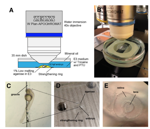
Set-up for time-lapse imaging of zebrafish lens epithelium (A) Schematic illustration of the imaging setup. (B) Real set-up picture. (C) The embryo was mounted in 1% low-melting agarose, and the yolk was put into the groove in order to orient the embryo laterally. (D) The agarose was restricted within the inner circle of a strengthening ring on the groove stretched into the acrylic plate. (E) The lateral side of the embryonic eye was used for confocal z-stack scanning. |
Expression Data
Expression Detail
Antibody Labeling
Phenotype Data
Phenotype Detail
Acknowledgments
This image is the copyrighted work of the attributed author or publisher, and
ZFIN has permission only to display this image to its users.
Additional permissions should be obtained from the applicable author or publisher of the image.
Full text @ Development