
Fig. 4
- ID
- ZDB-FIG-180822-56
- Publication
- Gribble et al., 2018 - The synaptic receptor Lrp4 promotes peripheral nerve regeneration
- Other Figures
- All Figure Page
- Back to All Figure Page
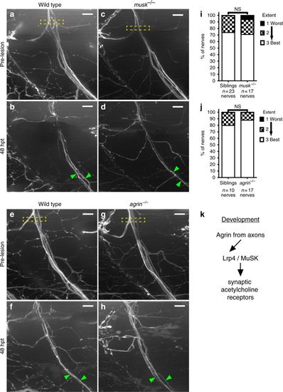
lrp4 acts in a MuSK-independent pathway to promote axonal regeneration. a Lateral view of wild-type GFP-labeled motor nerve before laser transection at 5 dpf. Yellow dashed box indicates transection site, scale bar is 10?�m. b Forty-eight hours post transection, multiple axon fascicles have regenerated ventrally. Green arrowheads indicate regenerated fascicles. c Lateral view of musk mutant motor nerve before laser transection at 5 dpf. Yellow dashed box indicates transection site. d Forty-eight hours post transection, motor axons have regenerated ventrally. Green arrowheads indicate regenerated fascicles. e Lateral view of wild-type GFP-labeled motor nerve before transection. f Forty-eight hours post transection, multiple axon fascicles regenerate ventrally. Green arrowheads mark regenerated fascicles. g Lateral view of agrin mutant motor nerve before transection at 5 dpf. Yellow box indicates transection site. h Forty-eight hours post transection, agrin mutant motor axons have regenerated ventrally. Green arrowheads mark regenerated fascicles. i Quantification of nerve regeneration in wild-type siblings (n?=?23 nerves from 11 larvae) and musk mutants (n?=?17 nerves from 11 larvae) at 48?h post transection. musk mutant motor nerves regenerate as well as wild-type sibling motor nerves (Fisher?s exact test p?=?0.4982). j Quantification of nerve regeneration in wild-type siblings (n?=?10 nerves from 4 larvae) and agrin mutants (n?=?17 nerves from 6 larvae). Agrin mutant motor nerves regenerate as well as wild-type sibling nerves (Fisher?s exact test p?=?0.6125). k Schematic of the canonical Agrin-Lrp4-MuSK pathway critical for neuromuscular synapse formation |
| Fish: | |
|---|---|
| Condition: | |
| Observed In: | |
| Stage Range: | Day 5 to Days 7-13 |