
Fig. 1
- ID
- ZDB-FIG-180822-53
- Publication
- Gribble et al., 2018 - The synaptic receptor Lrp4 promotes peripheral nerve regeneration
- Other Figures
- All Figure Page
- Back to All Figure Page
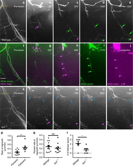
Pioneering axons establish regenerative path for follower axons. a?e Images from time lapse movie showing early regeneration of wild-type motor nerve with all axons expressing GFP. a Pre-lesion image; yellow box marks transection site. b A pioneering axon (magenta arrowhead and ?P?) crosses the injury gap ~800?min post-injury and extends ventrally. c, d, e Follower axons fasciculate with pioneering axon (numbered green arrowheads). f?j Still images from time lapse movie showing early regeneration of 5 dpf wild-type motor nerve; all axons express GFP, single-axon expresses mKate (magenta). f Pre-lesion image; yellow box marks transection site. Scale bar is 10?�m. g Axon sprouts in proximal nerve stump start extending and retracting ~400?min post transection. h Single-pioneering axon (magenta) crosses the injury gap and extends ventrally along distal nerve by 800?min post transection. Magenta arrowheads mark pioneer axon, which grows at a rate of 0.27?�m/min. i Follower axon extends and grows along pioneering axon; green arrowheads mark follower path, which grows at a rate of 0.36?�m/min. j By the end of imaging, the magenta pioneer axon has extended ventrally, as shown via a maximum projection image across time (magenta arrowheads). k?o Still images from time lapse movie showing early regeneration of lrp4 mutant motor nerve labeled with GFP. k Pre-lesion image; yellow box marks transection site. l Early growth cones sprout from proximal stump (open blue arrowheads) and pioneer axon extends ventrally ~600?min post transection (magenta arrowhead and ?P?). m?o Throughout imaging, only the pioneer axon extends to ventral myotome; follower axons explore transection site but do not traverse injury gap (open blue arrowheads). p Quantification of pioneer axon growth rate and follower axon growth rate in wild-type siblings. Pioneer axon average rate of regrowth is 0.24?�m/min (n?=?12 nerves in 8 larvae); follower axon average rate of regrowth is 0.47?�m/min (n?=?7 nerves in 4 larvae; unpaired t-test p?=?0.0055; t?=?3.181, df?=?17; error bars show mean and SEM). q Quantification of pioneer axon rate of regrowth in siblings and lrp4 mutants. Pioneer axons grow at equivalent rates: sibling average rate of regrowth?=?0.24?�m/min (n?=?12 nerves in 8 larvae); lrp4?/? average rate of regrowth?=?0.21?�m/min (n?=?10 nerves in 4 larvae; unpaired two-tailed t-test p?=?0.4702, t?=?0.7361, df?=?20; error bars show mean and SEM). r Number of follower axons in siblings and lrp4 mutants. In siblings, multiple follower axons extend (average number of follower axons?=?2.2, n?=?6 nerves in 2 larvae) while in lrp4 mutants, follower axons fail to extend ventrally (lrp4 mutant average number of follower axons?=?0.7, n?=?7 nerves in 2 larvae). Number of follower axons in siblings versus mutants is significantly different (two-tailed t-test p?=?0.0206, t?=?2.703, df?=?11; error bars show mean and SEM) |
| Fish: | |
|---|---|
| Condition: | |
| Observed In: | |
| Stage: | Day 5 |