
Fig. 4
- ID
- ZDB-FIG-180821-16
- Publication
- McMillan et al., 2018 - A regulatory pathway involving retinoic acid and calcineurin demarcates and maintains joint cells and osteoblasts in the fin regenerate
- Other Figures
- All Figure Page
- Back to All Figure Page
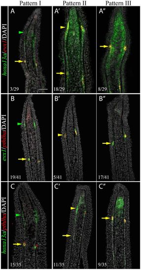
Sequential activation of hoxa13a, evx1 and pthlha expression in the presumptive joint cells. Double FISH and DAPI counterstains on longitudinal cryosections of 4?dpa fin regenerates illustrate three distinct patterns of expression. (A-C?) Patterns I and II are observed in rays with presumptive joint cells (arrowheads). (A?-C?) Pattern III is observed in rays without presumptive joint cells. (A-C?) In joint-forming cells (yellow arrows), the three markers are co-expressed in the joint-forming cells. (A,A?,C,C?) In the presumptive joint cells (green or yellow arrowheads), hoxa13a is expressed alone (Pattern I, A,C) or co-expressed with evx1 (Pattern II, A?) or pthlha (Pattern II, C?). (B?,C?) pthlha is also co-expressed with evx1 (B?). (B,B?) evx1 is either expressed alone (B) or co-expressed with pthlha (B?). Numbers in each panel represent the number of sections with the expression pattern over the total number of sections analyzed. Scale bar: 50??m (in A for A-C?). |
| Genes: | |
|---|---|
| Fish: | |
| Condition: | |
| Anatomical Term: | |
| Stage: | Adult |