Fig. 5
- ID
- ZDB-FIG-180621-73
- Publication
- Suniaga et al., 2018 - Increased mechanical loading through controlled swimming exercise induces bone formation and mineralization in adult zebrafish
- Other Figures
- All Figure Page
- Back to All Figure Page
|
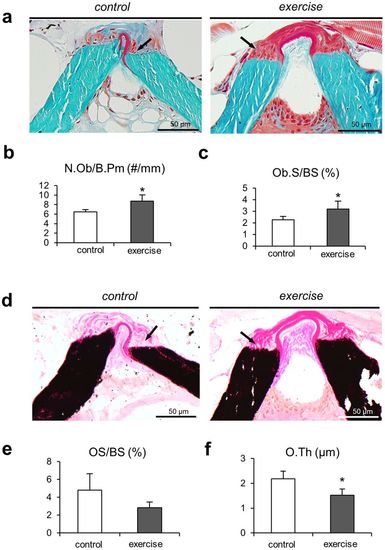
Bone formation mechanism. Static histomorphometric analysis of the bone formation mechanism in zebrafish. (a) Masson-Goldner trichrome-stained sections show the end plates of two adjacent vertebrae. Numerous osteoblasts (black arrows) can be observed in the same regions where fluorescence microscopy revealed increased bone formation. (b) Quantification of osteoblasts performed on Masson-Goldner trichrome-stained sections yielded higher osteoblast numbers and (c) greater osteoblast surface per bone surface in zebrafish from the exercise group. These results are consistent with higher bone deposition rates and higher bone volume found in the exercise group, given that pronounced osteoblast activity is associated with the deposition of new bone material. (d) Von Kossa/van Gieson stained sections display non-mineralized bone matrix, i.e. osteoid (black arrows), predominantly on the vertebral edges. (e) Osteoid surface per bone surface (OS/BS) remained similar between the groups. (f) Osteoid thickness (O.Th) was significantly lower in the exercise group compared to controls. |

