
Fig. 3
- ID
- ZDB-FIG-180621-71
- Publication
- Suniaga et al., 2018 - Increased mechanical loading through controlled swimming exercise induces bone formation and mineralization in adult zebrafish
- Other Figures
- All Figure Page
- Back to All Figure Page
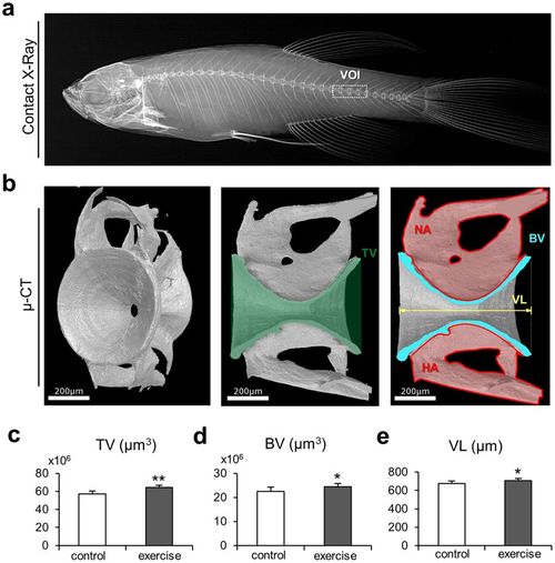
Bone microstructural changes due to musculoskeletal exercise. (a) Contact X-ray image displays the volume of interest (VOI) in the caudal spine of zebrafish. All parameters were extracted from caudal vertebrae. (b) Micro-CT images display the typical hourglass shape of the autocentrum that represents the core of the zebrafish vertebral body. The volume parameters extracted to quantify skeletal changes due to the swimming exercise are displayed on the sagittal cross-sections of the vertebrae: tissue volume (TV), bone volume (BV), and vertebral length (VL). The neural arch (NA) and the hemal arch (HA) were not included in the quantification of structural parameters via micro-CT. (c) Tissue volume was significantly higher in the exercise group. (d) Bone volume of the vertebral body was significantly greater in the exercise group. (e) Vertebral length increased substantially in the exercise group. The larger bone anatomy of the vertebral bodies of the zebrafish exposed to exercise indicates an increase in bone mass due to increased swimming activity. |