
Fig. 4
- ID
- ZDB-FIG-180621-72
- Publication
- Suniaga et al., 2018 - Increased mechanical loading through controlled swimming exercise induces bone formation and mineralization in adult zebrafish
- Other Figures
- All Figure Page
- Back to All Figure Page
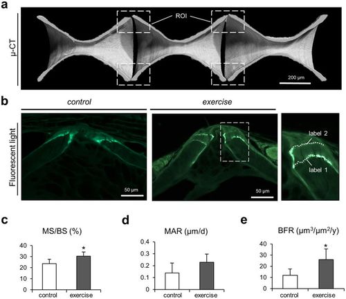
Characterization of bone formation by dynamic histomorphometry. (a) Micro-CT images of caudal vertebral bodies display the regions of interest (ROI) including the vertebral body end plates. (b) Fluorescence microscopy reveals fluorescent labeling in the end plates from both zebrafish groups. Larger inter-label distance can be observed in exercised zebrafish indicating higher bone formation activity. (c) Mineralized surface per bone surface (MS/BS) was significantly higher in the exercise group. (d) Mineral apposition rate (MAR) did not vary significantly between groups. (e) Bone formation rate (BFR) was significantly higher in exercised zebrafish. The increased deposition of new bone in the end plates of exercised zebrafish explains the increased vertebral length, bone volume and tissue volume measured using micro-CT (see Fig. 3). |