
Fig. 3
- ID
- ZDB-FIG-170815-16
- Publication
- Lim et al., 2017 - Caveolae Protect Notochord Cells against Catastrophic Mechanical Failure during Development
- Other Figures
- All Figure Page
- Back to All Figure Page
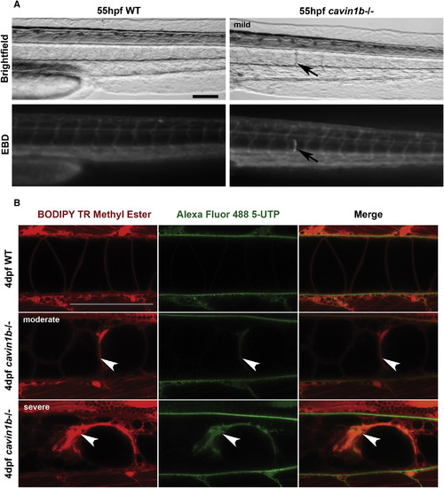
Notochord Lesions Are Damaged and Membrane-Permeable Cells (A) Evans Blue Dye (EBD) infiltration into membrane-compromised notochord cells in live 55-hpf embryos. Arrows mark the mild lesion with corresponding specific EBD uptake. Note no uptake in skeletal muscles. Four fish per group are shown. The scale bar represents 100 ?m. (B) Alexa 488 5-UTP infiltration into membrane-compromised notochord cells in live 4-dpf embryos. Colocalization of Alexa 488 5-UTP with BODIPY TR methyl ester (endomembranes) occurred in moderate (middle row) and severe lesions (bottom row). No uptake in skeletal muscles was observed. Arrowheads mark lesions. n = 5 (WT) and n = 10 (mutant). The scale bar represents 100 ?m. |
| Fish: | |
|---|---|
| Observed In: | |
| Stage Range: | Long-pec to Day 4 |