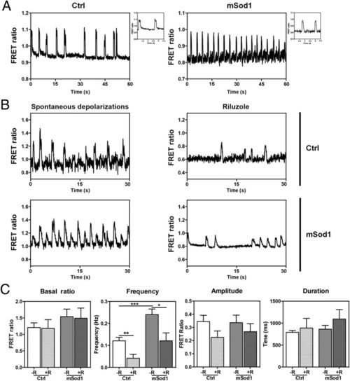
Riluzole treatment reduces spontaneous high-frequency depolarizations in the spinal motor neurons of mSod1 embryos.
(A) Representative examples of spontaneous mean FRET ratio changes in a control (Ctrl) and mSod1 motor neuron during a one-minute recording (the biosensor FRET ratio increases when membrane potential increases). Insets: detailed morphology of two depolarizing events. (B) Representative traces showing the FRET ratio changes recorded in the same Ctrl and mSod1 motor neurons before and after five minutes? riluzole administration. (C) In comparison with Ctrl, the mSod1 motor neurons showed a significant increase in the frequency of spontaneous depolarizations (Ctrl: 0.12?�?0.02?Hz; mSod1: 0.24?�?0.03?Hz), but there was no difference in the basal FRET ratio (Ctrl: 1.21?�?0.14; mSod1: 1.54?�?0.23), or the amplitude (Ctrl: 0.34?�?0.05; mSod1: 0.34?�?0.06) or duration of depolarization (Ctrl: 791?�?44?ms; mSod1: 863?�?86?ms). The statistical analyses showed that riluzole did not affect the motor neurons? basal FRET ratio (Ctrl: 1.18?�?0.26; mSod1: 1.49?�?0.31), or the amplitude (0.22?�?0.05 in Ctrl; mSod1 0.27?�?0.06) or duration of periodic depolarizations (Ctrl: 889?�?219?ms; mSod1: 1094?�?215?ms), but significantly reduced the frequency of the depolarizing events (Ctrl 0.04?�?0.02?Hz; mSod1 0.12?�?0.02?Hz). The columns in each graph represent the mean value?�?SEM of the indicated parameter in 12 Ctrl and 12 mSod1 motor neurons before and after riluzole administration. The measures were statistically analyzed using an unpaired Student t-test (*P?
|
