
Fig. S2
- ID
- ZDB-FIG-170209-20
- Publication
- Sotolongo-Lopez et al., 2016 - Genetic Dissection of Dual Roles for the Transcription Factor six7 in Photoreceptor Development and Patterning in Zebrafish
- Other Figures
- All Figure Page
- Back to All Figure Page
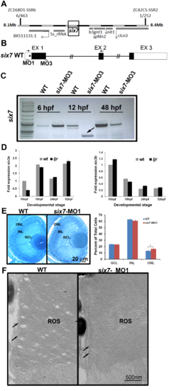
six3a/b expression is unchanged in six7 morphants. (A) Linkage analysis places the ljrp23ahub locus on chromosome 7, 6 out of 463 larvae show recombination at marker ZC168D1-SSR6 and 1 out of 252 larvae show recombination at marker ZC42C5-SSR2. (B) Diagram of predicted morpholino recognition sites (bars) in six7 loci. MO1 targets a translational site and MO3 blocks a donor splice site in intron 1 of six7. Incorrect splicing can be seen in morpholino-injected animals using primers in exon1 and 2 (arrows). (C) RT-PCR fragments, using primers highlighted in A, were analyzed by 1% agarose gel electrophoresis. Arrow highlights the six7 alternative spliced product obtained at 12 hpf (* indicates the new cryptic splice site). (D) No changes in the expression of the homologues six3a (left graph) or six3b (right graph) were detected between WT and ljrp23ahub mutants (n = 30 embryos per group). All the real-time PCR experiments were carried out in triplicates and normalized to ?-actin. (E) Plastic sections of 4 dpf retinas from WT and six7-MO1 injected larvae. The three layers are regularly arranged in WT and morphant retinas. Close examination of the retina revealed a densely packed ONL in morphants. Graph showing the average number of nuclei per unit area (WT, n = 3, 2 sections each; six7-MO1, n = 3, 2 sections each). Cells are increased in the ONL of six7 morphants. Student t test, arcsine transformation, *p<0.05. (F) Electron micrographs of rod outer segments (ROS) from central retina of WT and six7-MO1 larvae. ROS from six7 morphants show their typical morphology of parallel- flattened sacs with continuously membranes at the edges (arrows) indistinguishable from WT ROS. |