
Fig. 5
- ID
- ZDB-FIG-170201-19
- Publication
- Liu et al., 2016 - The Machado-Joseph Disease Deubiquitinase Ataxin-3 Regulates the Stability and Apoptotic Function of p53
- Other Figures
- All Figure Page
- Back to All Figure Page
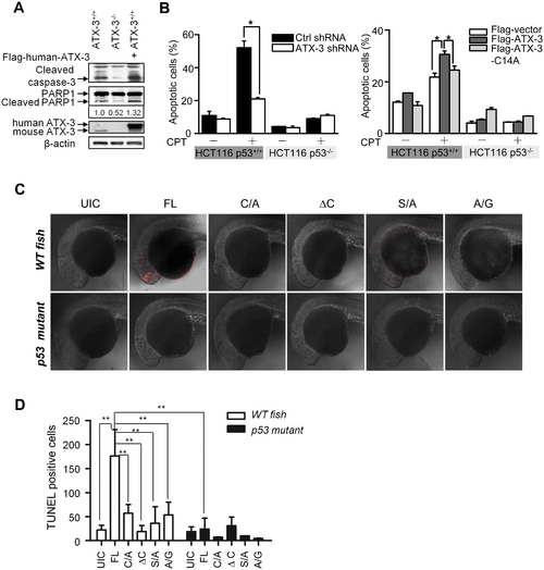
ATX-3 promotes p53-dependent apoptosis in cells and in zebrafish. (A) Western blot analysis of the involvement of ATX-3 in apoptosis as measured by the detection of caspase-3 and PARP1 cleavage in ATX-3+/+ and ATX-3-/- MEF cells, transfected with empty vector or plasmid encoding Flag-ATX-3. The relative changing fold of the cleaved PARP1 was underlined. (B) HCT116 p53+/+ and HCT116 p53-/- cells, stably shRNA-expressing cells, or cells transiently transfected with Flag-vector or Flag-ATX-3 or Flag-ATX-3-C14A were treated with or without CPT (1 μM) for 24 h. Cells were analyzed by flow cytometry for apoptosis using Annexin V/PI. Results are the mean ± SEM of three independent experiments. * denotes p < 0.05. Underlying data are shown in S1 Data. (C) WT and p53 mutant zebrafish embryos were injected with indicated object mRNAs (100 pg) at the one-cell stage and harvested at 24 h post fertilization (hpf) for TUNEL labeling and observed by confocal microscopy. Embryos were shown in lateral view with anterior to the left. UIC, uninjected control. (D) Quantitative analysis of Fig 5C. The data represent the mean ± SEM for TUNEL-positive cells in 3~9 zebrafish embryos. ** denotes p < 0.01. Underlying data are shown in S1 Data. |