
Fig. S3
- ID
- ZDB-FIG-161122-44
- Publication
- Mouti et al., 2016 - Minimal contribution of ERK1/2-MAPK signalling towards the maintenance of oncogenic GNAQQ209P-driven uveal melanomas in zebrafish
- Other Figures
- All Figure Page
- Back to All Figure Page
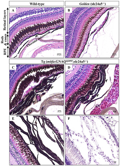
Choroidal hyperplasia is evident in pigmentation-rescued Tg (mitfa:GNAQQ209P;slc24a5-/-) zebrafish. A-D. H&E staining of transverse sections of formalin-fixed and paraffin-embedded eye tissues of a 5-month-old adult zebrafish. (A) Wild-type demonstrating different structures of the zebrafish retina as indicated. (B) Golden mutant: note the hypopigmented RPE and structurally normal choroid. (C, D) Tg (mitfa:GNAQQ209P;slc24a5-/-): note here benign hyperplastic choroidal lesions (black arrowheads) developing beneath pigmentation-rescued RPE. E. A representative example of H&E stained transverse section illustrating thickened and heavily pigmented choroidal melanocyte layer before melanin bleaching. F. Multiple cellular layers of thickened choroid, as revealed by multiple nuclei (blue; counterstained with hematoxylin) after melanin bleaching. Abbreviations: RPE, retinal pigmented epithelium. Scale bar lengths, as indicated. |