Fig. 5
- ID
- ZDB-FIG-160729-7
- Publication
- Lv et al., 2016 - Synaptic Ribbons Require Ribeye for Electron Density, Proper Synaptic Localization, and Recruitment of Calcium Channels
- Other Figures
- All Figure Page
- Back to All Figure Page
|
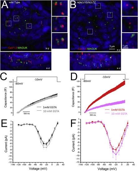
re[a(Δ10)/b(Δ7)] Homozygous Hair Cells Have Poorly Localized Calcium Channels and Exhibit Enhanced Sensitivity to 10 mM EGTA (A and B) Immunolabel of CaV1.3a and MAGUK in 5-dpf WT (A) and re[a(Δ10)/b(Δ7)] (B). While Cav1.3 immunolabeled puncta generally localize to the presynapse adjacent to postsynaptic MAGUK immunolabel in WT (A; insets), CaV1.3a clusters fail to localize to the presynapse in re[a(Δ10/b(Δ7)] mutants (B; inset). (C and D) Capacitance increase of WT and double-mutant neuromast hair cells in response to a 3-s step depolarization with 1 mM EGTA or 10 mM EGTA (gray, n = 8) in the internal solution. The average capacitance increase at the end of the 3-s depolarization (averaged over 100 ms) of WT fish was 67.8 ± 5.0 fF at 1 mM EGTA (C; black, n = 11) and 62.8 ± 4.9 fF at 10 mM EGTA (C; gray, n = 7); p = 0.50. 10 mM EGTA blocks 7.4% of release, compared to 1 mM EGTA in WT fish. The average capacitance increase at the end of the 3-s depolarization (averaged over 100 ms) of double mutants was 106.1 ± 13.6 fF at 1 mM EGTA (D; red, n = 12), and 36.3 ± 8.7 fF at10 mM EGTA (D; pink, n = 8), p = 0.01. 10 mM EGTA blocks 66.% of release compared to 1 mM EGTA. (E and F), Plot of current-voltage relationship for WT fish NM hair cells (E) and re [a(Δ10)/b(Δ7)] homozygous mutant neuromast hair cells (F) at 1 mM EGTA and 10 mM EGTA internal solution. At 20 mV, the average current of WT cells was 6.1 ± 0.5 pA at 1 mM EGTA (E; red, n = 15) and 6.9 ± 0.9 (E; pink, n = 8). p = 0.46. The average current of mutant fish cells was 8.26 ± 0.58 pA at 1 mM EGTA solution (F; red, n = 17), and 9.9 ± 2.2 pA at 10 mM EGTA solution (F; pink, n = 6). p = 0.49. The fish recorded were between 5 and 8 days old, and the neuromasts recorded were P3 and P4. |
| Gene: | |
|---|---|
| Antibodies: | |
| Fish: | |
| Anatomical Terms: | |
| Stage: | Day 5 |
| Fish: | |
|---|---|
| Observed In: | |
| Stage: | Day 5 |

