
Fig. 1
- ID
- ZDB-FIG-160714-16
- Publication
- Richardson et al., 2016 - Re-epithelialization of cutaneous wounds in adult zebrafish uses a combination of mechanisms at play during wound closure in embryonic and adult mammals
- Other Figures
- All Figure Page
- Back to All Figure Page
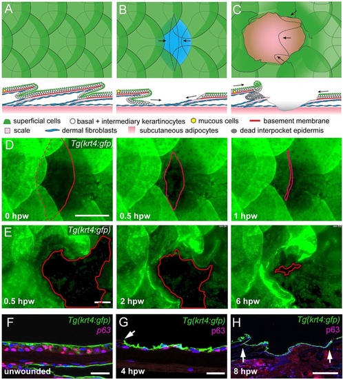
Re-epithelialization of full- and partial-thickness wounds in adult zebrafish. (A-C) Schematic representations of normal adult zebrafish skin architecture (A), or following partial-thickness (B) or full-thickness (C) wound introduction. Arrows in B and C indicate the typical directions of epidermal re-epithelialization. (D,E) Single images from time-lapse recordings of Tg(krt4:GFP) fish following partial-thickness (D) or full-thickness (E) wounding at indicated time points post-wounding; red line marks LE. (F-H) Unwounded epidermis is 3-4 cell layers thick (F), the re-epithelializing neo-epidermis is bilayered, consisting of superficial GFP+ and inner p63+ keratinocytes (G,H). Arrow in G indicates LE, arrows in H, the wound margins, revealing full re-epithelialization at 8hpw. Scale bars: 500�m in D,E,H; 20�m in F,G. |
| Gene: | |
|---|---|
| Antibody: | |
| Fish: | |
| Conditions: | |
| Anatomical Terms: | |
| Stage: | Adult |