
|
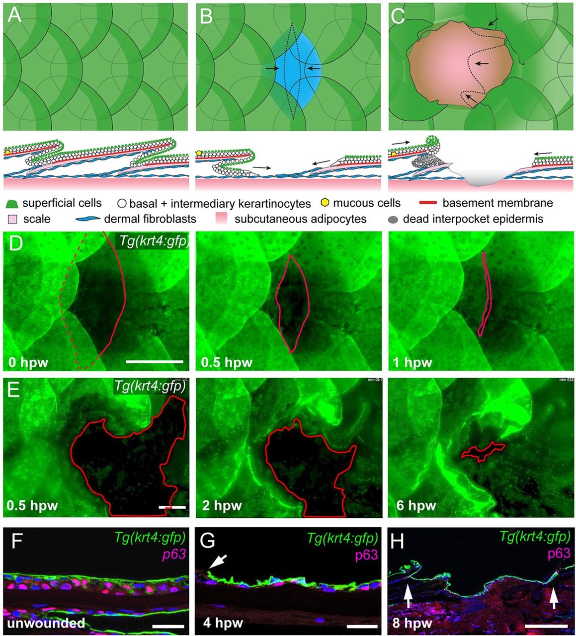
Fig. 1
Re-epithelialization of full- and partial-thickness wounds in adult zebrafish. (A-C) Schematic representations of normal adult zebrafish skin architecture (A), or following partial-thickness (B) or full-thickness (C) wound introduction. Arrows in B and C indicate the typical directions of epidermal re-epithelialization. (D,E) Single images from time-lapse recordings of Tg(krt4:GFP) fish following partial-thickness (D) or full-thickness (E) wounding at indicated time points post-wounding; red line marks LE. (F-H) Unwounded epidermis is 3-4 cell layers thick (F), the re-epithelializing neo-epidermis is bilayered, consisting of superficial GFP+ and inner p63+ keratinocytes (G,H). Arrow in G indicates LE, arrows in H, the wound margins, revealing full re-epithelialization at 8hpw. Scale bars: 500�m in D,E,H; 20�m in F,G.