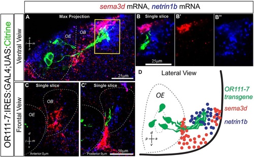
Fig. 1
- ID
- ZDB-FIG-160218-8
- Publication
- Taku et al., 2016 - Attractant and repellent cues cooperate in guiding a subset of olfactory sensory axons to a well-defined protoglomerular target
- Other Figures
- All Figure Page
- Back to All Figure Page
|
sema3d and netrin 1b have complementary expression patterns in the zebrafish OB at 36hpf. (A) Maximum intensity projection spanning 30µm through a 36hpf or111-7:IRES:GAL4:UAS:citrine embryo. Ventral view, anterior is up and medial is to the right. Axons are shown in green, sema3d mRNA in red and netrin 1b mRNA in blue. (B-B′′) A single optical section through the inset in A. (C,C′) Single optical sections through a 36hpf embryo. Frontal view, dorsal is up and medial is to the right. Sections are arranged from anterior (left) to posterior (right). The distance from each section to the anteriormost part of the telencephalon is denoted in bottom left. (D) Diagram of a 36hpf embryo in lateral view. sema3d is expressed in the anterior OB. Some netrin 1b is detected in the anterior bulb but it extends further posteriorly. or111-7 transgene-expressing axons are positioned posterior to sema3d expression but within the netrin 1b expression domain. or111-7 transgenic axons are not present in the anteriormost portion of the telencephalon. sema3d expression wraps around the edge of the olfactory pit and is also present between the OE and nascent OB. OE, olfactory epithelium; OB, olfactory bulb. |
| Genes: | |
|---|---|
| Fish: | |
| Anatomical Terms: | |
| Stage Range: | Prim-25 to High-pec |

