
Fig. 2
- ID
- ZDB-FIG-151221-4
- Publication
- Chua et al., 2015 - Tumor-specific signaling to p53 is mimicked by Mdm2 inactivation in zebrafish: insights from mdm2 and mdm4 mutant zebrafish
- Other Figures
- All Figure Page
- Back to All Figure Page
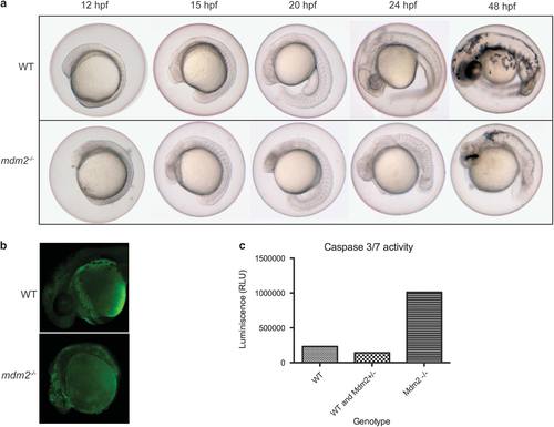
Embryonic development and analysis of cell death in wild-type and mdm2-/- embryos. Embryos were obtained from an incross of wild-type or mdm2+/- zebrafish. (a) Brightfield images of wild-type and mdm2-/-embryos at matching developmental stages show the lethal phenotype due to the homozygous loss of mdm2. (b) The 24-hpf wild-type and mdm2-/- embryos were stained live with acridine orange and imaged under ultraviloet light. Increased green fluorescence in mdm2-/- embryos compared with wild-type embryos suggests the presence of more apoptotic cells. (c) Whole-cell lysate was prepared from a number of wild-type/mdm2+/- and mdm2-/- embryos and used in a Caspase 3/7 assay. Cells from mdm2-/- embryos have higher Caspase 3/7 activity compared with wild-type/mdm2+/- siblings. |
| Fish: | |
|---|---|
| Observed In: | |
| Stage Range: | 5-9 somites to Long-pec |