Fig. 2
- ID
- ZDB-FIG-151123-5
- Publication
- Hamling et al., 2015 - Mapping the development of cerebellar purkinje cells in zebrafish
- Other Figures
- All Figure Page
- Back to All Figure Page
|
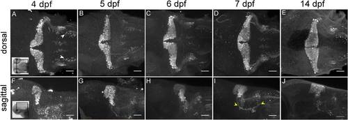
Parvalbumin7 expression reveals morphological changes in the Purkinje cell layer between 4 and 14 dpf. Maximum intensity projections of Pvalb7 expression from dorsal (A–E) and sagittal (F–I) views in 4–14 dpf larvae. Insets represent approximate dorsal and sagittal brain region imaged (A, F). The wing-shaped morphology of the Purkinje cell layer is present at all developmental time points (A–E), though it begins to wrap more anteriorly (see G–I) and Purkinje cells begin to project cerebellovestibular axons (yellow arrowheads) by 7 dpf (I). Dorsolateral hindbrain neurons are present by 4 dpf (A, white arrowheads). Scale bar 50 µm. Images taken using 20× (1.0 NA) water objective. |
| Antibody: | |
|---|---|
| Fish: | |
| Anatomical Term: | |
| Stage Range: | Day 4 to Days 14-20 |

