
Fig. 4
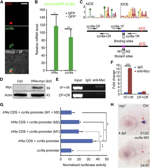
ccr9a Is a Direct Target of Irf4a (A) Co-expression of irf4a and ccr9a in the CHT of 2-dpf embryos revealed by double color FISH. Scale bar, 100 �m. (B) qPCR of irf4a and ccr9a in GFP+ cells relative to negative cells sorted from coro1a:EGFP transgenic embryos at 2 dpf. p < 0.05; p < 0.01. (C) Schematic diagram of the ChIP and reporter assay. The ccr9a-CF and CR are the control forward and reverse primers, which are used to amplify the ccr9a promoter region without the conserved Irf4 binding site. The ccr9a-1F and 1R represent primers used to amplify the promoter region with two conserved Irf4 binding sites within 500 bp upstream of 5′-ATG-3′. AICE, AP-1-IRF composite elements; EICE, Ets-IRF composite elements. (D) Myc was detected in embryos injected with irf4a-myc mRNA by western blot. (E) Direct binding of ccr9a promoter by Irf4a examined by ChIP assay. (F) qPCR of DNA fragments obtained from ChIP assay with primers (ccr9a-1F, 1R or CF, CR). p < 0.001. (G) Reporter assay showed the regulation of ccr9a promoter by Irf4a full-length CDS. M1 indicates that 5′-TGAT-3′ within AICE was mutated to 5′-GTCG-3′, and M2 indicates that 5′-GGAA-3′ within EICE was mutated to 5′-TTCC-3′. p < 0.01; p < 0.05. (H) Expression of rag1 in the thymus (red arrow) of the control and ccr9a morphants at 4 dpf. All data are mean � SD. See also Figure S4 and Table S2. |
| Genes: | |
|---|---|
| Fish: | |
| Knockdown Reagent: | |
| Anatomical Terms: | |
| Stage Range: | Long-pec to Day 4 |
| Fish: | |
|---|---|
| Knockdown Reagent: | |
| Observed In: | |
| Stage: | Day 4 |
Reprinted from Developmental Cell, 34(6), Wang, S., He, Q., Ma, D., Xue, Y., Liu, F., Irf4 Regulates the Choice between T Lymphoid-Primed Progenitor and Myeloid Lineage Fates during Embryogenesis, 621-31, Copyright (2015) with permission from Elsevier. Full text @ Dev. Cell