
Fig. 3
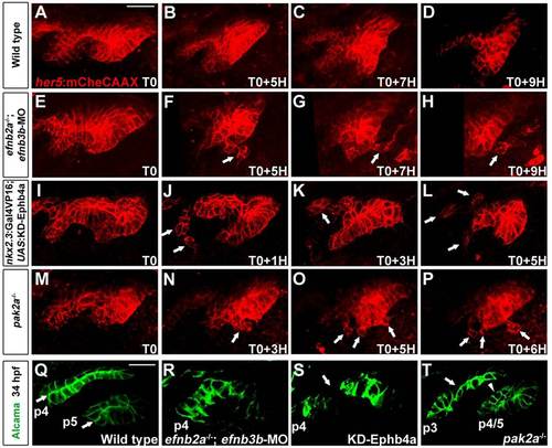
Pouch disintegration in the absence of EphB-Pak2a signaling. (A-P) Confocal projections from time-lapse recordings show development of her5:mCherryCAAX+ pouches p4-p6 (red). In embryos with reduced Efnb2a/b3b, endoderm-specific expression of KD-Ephb4a or mutant for pak2a, cells break off from the epithelium (arrows) as pouches migrate outwards. Movies start at 25hpf (T0). (Q-T) Confocal sections show pouches labeled by Alcama immunohistochemistry at 34hpf. Whereas wild-type pouch epithelial cells form bilayers with a straight line of apical membranes (arrows in Q), apical membranes are variably disorganized in embryos with reduced Efnb2a/b3b, endoderm-specific expression of KD-Ephb4a or mutant for pak2a. Note dissociated pouch epithelial cells (arrow) in nkx2.3:Gal4VP16; UAS:KD-Ephb4a embryos (S) and regions of monolayered (arrow) and multilayered (arrowhead) epithelia in pak2a mutant pouches (T). Scale bars: 20�m. |