Fig. 2
- ID
- ZDB-FIG-150330-31
- Publication
- Fukui et al., 2014 - S1P-Yap1 Signaling Regulates Endoderm Formation Required for Cardiac Precursor Cell Migration in Zebrafish
- Other Figures
- All Figure Page
- Back to All Figure Page
|
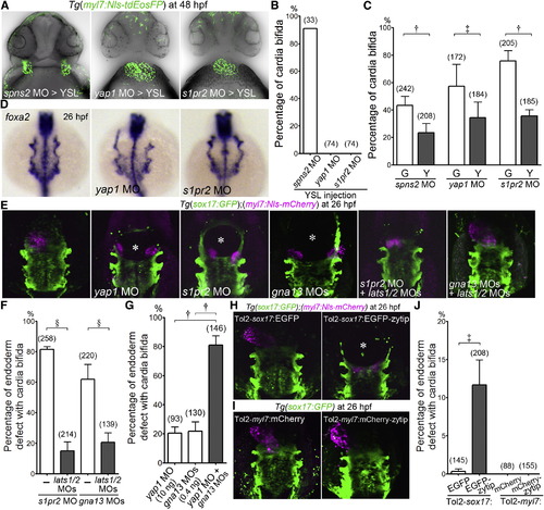
Yap1 Functions Downstream of S1P-S1pr2 Signaling in the Endoderm (A) Confocal images of Tg(myl7:Nls-tdEosFP) embryos at 48 hpf injected with MO indicated at the bottom into the yolk syncytial layer (YSL). (B) Quantitative analyses of the incidence of cardia bifida of (A). Graph shows the percentage of the embryos with cardia bifida among the total embryos injected with MO indicated at the bottom. (C) The incidence of cardia bifida of Tg(myl7:Nls-tdEosFP) embryos injected with MO and the plasmids (30 pg/embryo) transiently expressing either EGFP (G) or EGFP-tagged Lats1/2 kinase-insensitive Yap1 (Y) under the control of sox17 promoter using Tol2 transposon system. (D) WISH analyses of foxa2 expression of the embryos (26 hpf) uninjected (left) and injected with MO indicated at the bottom (center and right). Note that foxa2 mRNA was detected in the endoderm and in the notochord and that the space between bilateral pharyngeal endoderm was widened in the yap1 morphants and s1pr2 morphants. A set of representative images of four independent experiments is shown as dorsal views. (E) 3D-rendered two-photon laser-scanned z-stack images of Tg(sox17:GFP);(myl7:Nls-mCherry) embryos uninjected (left) and injected with MO indicated at the bottom. gna13 MO stands for both gna13a and gna13b MOs. Asterisks indicate the defect of the endoderm. (F and G) Quantitative analyses of incidence of the defects in the endoderm with cardia bifida in the embryos treated with the MOs indicated at the bottom. (H and I) Images of those injected with the plasmid indicated at the top (25 pg/embryo) into the embryo indicated at the top. Note that forced expression of zYtip in the endoderm resulted in the defect of the endoderm with cardia bifida while that in the CPCs did not. Asterisk indicates the endodermal defect. (J) Quantitative analyses of the results obtained in (H) and (I). §p < 0.0001, ?p < 0.01, ?p < 0.05. See also Figure S2. |
| Genes: | |
|---|---|
| Fish: | |
| Knockdown Reagents: | |
| Anatomical Terms: | |
| Stage: | Prim-5 |
Reprinted from Developmental Cell, 31, Fukui, H., Terai, K., Nakajima, H., Chiba, A., Fukuhara, S., Mochizuki, N., S1P-Yap1 Signaling Regulates Endoderm Formation Required for Cardiac Precursor Cell Migration in Zebrafish, 128-136, Copyright (2014) with permission from Elsevier. Full text @ Dev. Cell

