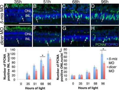
Dicer knockdown decreased INL proliferation in the light-damaged retina. Lissamine-tagged dicer 5-base mismatch (5-mis) control morpholino or dicer morpholino were intravitreally injected and electroporated into dark-adapted adult albino zebrafish. Retinas were collected at 0, 35, 51, 68, and 96h of light damage and immunostained with anti-PCNA (green) antibodies and TOPRO3 nuclear stain (blue). A, E: At 35h, single PCNA-positive M�ller glia were observed in the INL of dicer 5-mis control and dicer morphant retinas (arrows). B, F: Both dicer 5-mis control and dicer morphant retinas contain doublet nuclei at 51h of light damage (arrows). C: Clusters of proliferating progenitor cells are present in the INL of dicer 5-mis control retinas (arrow) at 68h of light damage. G: Single (arrow) or doublet (arrowhead) nuclei predominated in dicer morphant retinas. D: Columns of proliferating progenitors were observed in the INL of dicer 5-mis control morphant retinas at 96h of light damage. H: At 96h of light damage, doublet nuclei (arrowhead) predominated the INL of dicer morphants. I: Significantly fewer PCNA-positive INL cells were present in dicer morphant retinas compared to dicer 5-mis control morphant retinas beginning at 68h of light damage. J: dicer morphant retinas contained significantly fewer PCNA-positive ONL cells at 96h of light damage. dicer 5-mis MO, dicer 5-base mismatch control morphant; dicer MO, dicer morphant; INL, inner nuclear layer; ONL, outer nuclear layer. Scale bar in A = 50 �m and applies to B?H; data represent mean � s.e.m; *P< 0.05 using two-way ANOVA with a Tukey′s post-hoc test, n=11.
|
