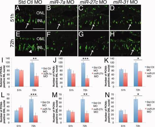
Knockdown of miR-7a, miR-27c and miR-31 reduced proliferation at 72h. Adult albino zebrafish were intravitreally injected and electroporated with either standard control miR-7a, miR-27c or miR-31 morpholino prior to the start of light damage. A?D: After 51h of light, standard control, miR-7a, miR-27c, and miR-31 morphant retinas contained PCNA-positive nuclei in the INL (arrows). E: At 72h, standard control morphant retinas contain columns of INL proliferating nuclei (arrowhead). F?H: At 72h, miR-7a, miR-27c and miR-31 morphant retinas contained mainly doublet INL and single nuclei (arrows). I?K: There were no significant differences in the number of PCNA-positive INL cells at 51h between the standard control morphant retina and the miR-7a (I), miR-27c (J), and miR-31 (K) morphant retinas. In contrast, significantly fewer PCNA-positive INL cells were present in all three miRNA morphant retinas relative to standard control morphant retinas at 72h. L?N: There were no significant differences in the number of PCNA-positive ONL cells at 51h between the standard control morphant retina and the miR-7a (L), miR-27c (M), and miR-31 (N) morphant retinas. In contrast, significantly fewer PCNA-positive INL cells were present in all three miRNA morphant retinas compared to standard control morphant retinas at 72h. INL, inner nuclear layer; ONL, outer nuclear layer. Scale bar in A = 50 �m and applies to B?H. Data represent mean � s.e.m. *P< 0.05; ** P < 0.01; *** P <0.001 using two-way ANOVA with Tukey′s post-hoc test, n=6.
|
