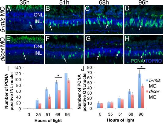
Fig. 2 Dicer knockdown decreased INL proliferation in the light-damaged retina. Lissamine-tagged dicer 5-base mismatch (5-mis) control morpholino or dicer morpholino were intravitreally injected and electroporated into dark-adapted adult albino zebrafish. Retinas were collected at 0, 35, 51, 68, and 96h of light damage and immunostained with anti-PCNA (green) antibodies and TOPRO3 nuclear stain (blue). A, E: At 35h, single PCNA-positive Müller glia were observed in the INL of dicer 5-mis control and dicer morphant retinas (arrows). B, F: Both dicer 5-mis control and dicer morphant retinas contain doublet nuclei at 51h of light damage (arrows). C: Clusters of proliferating progenitor cells are present in the INL of dicer 5-mis control retinas (arrow) at 68h of light damage. G: Single (arrow) or doublet (arrowhead) nuclei predominated in dicer morphant retinas. D: Columns of proliferating progenitors were observed in the INL of dicer 5-mis control morphant retinas at 96h of light damage. H: At 96h of light damage, doublet nuclei (arrowhead) predominated the INL of dicer morphants. I: Significantly fewer PCNA-positive INL cells were present in dicer morphant retinas compared to dicer 5-mis control morphant retinas beginning at 68h of light damage. J: dicer morphant retinas contained significantly fewer PCNA-positive ONL cells at 96h of light damage. dicer 5-mis MO, dicer 5-base mismatch control morphant; dicer MO, dicer morphant; INL, inner nuclear layer; ONL, outer nuclear layer. Scale bar in A = 50 µm and applies to B?H; data represent mean ± s.e.m; *P< 0.05 using two-way ANOVA with a Tukey′s post-hoc test, n=11.
Image
Figure Caption
Figure Data
Acknowledgments
This image is the copyrighted work of the attributed author or publisher, and
ZFIN has permission only to display this image to its users.
Additional permissions should be obtained from the applicable author or publisher of the image.
Full text @ Dev. Dyn.

