
Fig. 8
- ID
- ZDB-FIG-140917-46
- Publication
- Carr et al., 2014 - Characterization of the Zebrafish Homolog of Zipper Interacting Protein Kinase
- Other Figures
- All Figure Page
- Back to All Figure Page
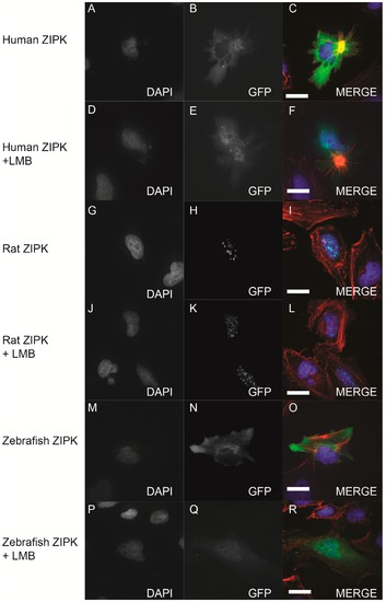
Nucleo-cytoplasmic shuttling of zebrafish ZIPK. HeLa cells were transfected with either human GFP-ZIPK (A?F); rat GFP-ZIPK (G?L); or zebrafish GFP-ZIPK (M?R). All cells were fixed and stained with DAPI and Alexa 568-phalloidin. The cells in (A?C, G?I, M?O) were treated with 0.1% DMSO; while the cells in (D?F, J?L, P?R) were treated with 50 nM leptomycin B for four hours. (A, D, G, J, M, P) are stained with DAPI; (B, E, H, K, N, Q) show subcellular localization of the GFP-fusion protein, while color images are a merge of DAPI (blue), GFP (green), and phalloidin (red). Each experiment was repeated three times with a minimum of 25 cells assayed per experiment. White bar indicates 20 μm. |