Fig. S2
- ID
- ZDB-FIG-140211-20
- Publication
- Dyer et al., 2014 - A bi-modal function of Wnt signalling directs an FGF activity gradient to spatially regulate neuronal differentiation in the midbrain
- Other Figures
- All Figure Page
- Back to All Figure Page
|
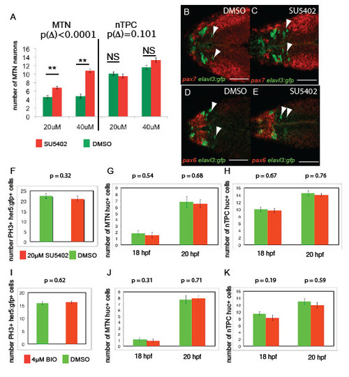
FGF and Wnt signalling regulates MTN number between 14-24 hpf, but not nTPC number, progenitor cell specification or proliferation. Treatment of embryos from 14 hpf with SU5402 at 20μM compared to 40μM results in a statistically significant increase in number (A, difference of change Δ following SU5402 treatment compared to DMSO, p Δ = 9.51x10⁻⁵, n=30). Transgenic Tg[elavl3:gfp] embryos exposed to 40μM SU5402 from 14 hpf (C) reveal that ectopic MTN neurons (green, anti-GFP) form within the pax7 expressing midbrain (red, pax7 in situ) at 24 hpf relative to DMSO treated controls (F), but are not forming in pax6+ forebrain regions (D,E). Quantification of proliferating cells in the midbrain of Tg[her5:gfp] embryos at 18 hpf following exposure to 20μM SU5402 (F) or 4μM BIO (I) from 14 hpf reveals no significant differences relative to DMSO treated control animals (p>0.05 for all, n=20). Quantification of huc/ elavl3 expressing neuronal progenitors in the midbrain or posterior diencephalon of 20 hpf and 18 hpf embryos treated with 20μM SU5402 (G,H) or 4μM BIO (J,K) from 14 hpf reveals no significant differences relative to DMSO treated control animals (p >0.05 for all, n=20). Error bars represent s.e.m., statistical comparisons were performed using unpaired t-tests. Scale bars 100μm 50μm (B-E). |

