Fig. 5
- ID
- ZDB-FIG-140211-16
- Publication
- Dyer et al., 2014 - A bi-modal function of Wnt signalling directs an FGF activity gradient to spatially regulate neuronal differentiation in the midbrain
- Other Figures
- All Figure Page
- Back to All Figure Page
|
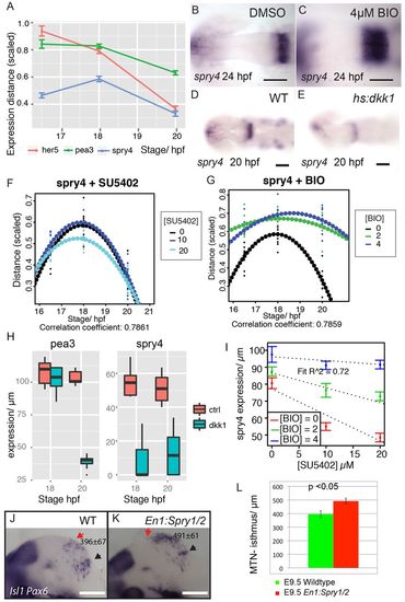
Wnt regulation of Sprouty genes controls FGF activity at the isthmus and directs neuronal positioning. Zebrafish pea3, her5 and spry4 midbrain expression between 16.5 and 20 hpf (A); distance is scaled by midbrain size; error bars indicate s.e.m., n=30. Expression of spry4 at 24 hpf in embryos treated from 14 hpf with DMSO (B) or 4 μM BIO (C) or spry4 at 20 hpf in Tg[hsp70:dkk1b-egfp] transgenic embryos (E) and siblings (D) after heat-shock at 16.5 hpf. Expression (scaled to midbrain size) of spry4 in the dorsal midbrain between 16.5 and 20 hpf after exposure to SU5402 (F) or BIO (G) from 14 hpf. Quadratic polynomials (dotted line) were fitted to plots of spry4 expression (μm) over time after DMSO, SU5402 or BIO exposure from 14 hpf at varying concentrations (n=10 for each condition). Plots of pea3 and spry4 expression at 18 and 20 hpf, following heat-shock induction of dkk1 at 16.5 hpf in Tg[hsp70:dkk1b-egfp] embryos and non-transgenic siblings (H). Plots of spry4 expression at 24 hpf after exposure to varying SU5402 and BIO concentrations from 14 hpf were fitted with lines; the decreasing slopes of expression as BIO concentration is increased reveals that BIO upregulates spry4 and attenuates an SU5402-induced reduction of spry4 expression (I); data are average with s.e.m. and the model agreement is reported by the R2 value for linear models (n=90). Expression of Isl1 and Pax6 in E9.5 wild-type (J) and En1:Spry1/2-/- (K) mice was quantified (L) to reveal that MTN neurons are anteriorly displaced after loss of Sprouty function in the midbrain; values represent distance (Ám) between the red arrow (MTN) and black arrow (isthmus); error bars indicate s.e.m. (n=10). Scale bars: 200 μm in J,K; 100 μm in D,E; 50 μm in B,C. |

