
Fig. 2
- ID
- ZDB-FIG-140131-23
- Publication
- Antinucci et al., 2013 - Teneurin-3 specifies morphological and functional connectivity of retinal ganglion cells in the vertebrate visual system
- Other Figures
- All Figure Page
- Back to All Figure Page
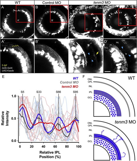
Teneurin-3 Is Required for Correct Stratification of RGC Dendrites (A–D) Kaede-expressing RGCs in the retina of 5 dpf WT, control MO-injected, and tenm3 MO-injected larvae. (A′–D′) Insets in (A)–(D) showing the dendritic stratification pattern of Kaede-positive RGCs. All images represent maximum intensity projections of ~20 μm confocal z stacks. Scale bars, 40 μm (A–D) and 20 μm in (A′–D′). GCL, ganglion cell layer; INL, inner nuclear layer; IPL, inner plexiform layer; OPL, outer plexiform layer. (E) Fluorescence profiles of IPL stratification in 5 dpf WT (blue), control MO-injected (gray), and tenm3 MO-injected (red) larvae. Thin traces represent intensity profiles of IPLs of single larvae. Thick traces indicate average profiles (WT, n = 7 larvae; control MO, n = 7; tenm3 MO, n = 10). Zero percent corresponds to the boundary between GCL and IPL, whereas 100% corresponds to the boundary between IPL and INL. (F) Schematic summarizing the defects observed in tenm3 morphant retinae. RGCs are indicated in blue. Neuropil layers are in gray. ONL, outer nuclear layer. See also Figure S4. |
| Gene: | |
|---|---|
| Fish: | |
| Knockdown Reagent: | |
| Anatomical Terms: | |
| Stage: | Day 5 |
| Fish: | |
|---|---|
| Knockdown Reagent: | |
| Observed In: | |
| Stage: | Day 5 |