
Fig. S3
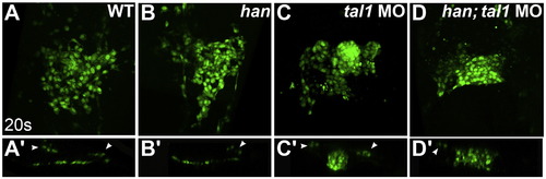
Endocardial aggregation in tal1-deficient embryos does not require hand2 function. (A?D) Three-dimensional reconstructions of confocal stacks depict dorsal views of wild-type (A), han mutant (B), tal1-deficient (C) and han mutant; tal1-deficient (D) embryos expressing Tg(fli1a:negfp) (green) at 20s. (A′?D′) Reconstructions of optical sections displaying transverse slices through the endocardium of the embryos shown in (A?D). Arrowheads mark progenitors of the lateral dorsal aortae. The endocardium forms a single-layered sheet in wild-type (A and A′) and han mutant (B and B′) embryos. In contrast, the endocardium in embryos that are deficient for both han and tal1 (D and D′) forms a multi-layered aggregate similar to that seen in tal1-deficient embryos (C and C′). |
Reprinted from Developmental Biology, 383(2), Schumacher, J.A., Bloomekatz, J., Garavito-Aguilar, Z.V., and Yelon, D., tal1 regulates the formation of intercellular junctions and the maintenance of identity in the endocardium, 214-226, Copyright (2013) with permission from Elsevier. Full text @ Dev. Biol.