
Fig. 4
- ID
- ZDB-FIG-131210-43
- Publication
- Peterson et al., 2013 - Dvr1 transfers left-right asymmetric signals from Kupffer's vesicle to lateral plate mesoderm in zebrafish
- Other Figures
- All Figure Page
- Back to All Figure Page
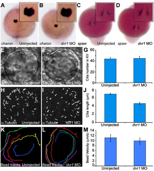
KV morphology and flow is normal in dvr1 morphants. (A) and (B) Cha expression in (A) uninjected embryos and (B) dvr1 morphants. (C) and (D) Peri-nodal spaw expression in (C) uninjected and (D) dvr1 morphant embryos. (E) and (F) KV shape in (E) uninjected and (F) dvr1 morphant embryos. (G) KV cilia number in wildtype and dvr1 morphant embryos. (H) and (I) KV cilia labeled by α-alpha-tubulin antibody in (G) uninjected and (H) dvr1 morphant embryos. (J) KV cilia length in wildtype and dvr1 morphant embryos. (K) and (L) Fluorescent bead tracking of fluid flow in KV in (K) uninjected wildtype and (L) dvr1 morphants. Each colored line represents the track of an individual bead. (M) Average fluorescent bead velocity. |
| Genes: | |
|---|---|
| Antibody: | |
| Fish: | |
| Knockdown Reagent: | |
| Anatomical Terms: | |
| Stage: | 5-9 somites |
| Fish: | |
|---|---|
| Knockdown Reagent: | |
| Observed In: | |
| Stage: | 5-9 somites |
Reprinted from Developmental Biology, 382(1), Peterson, A.G., Wang, X., and Yost, H. J., Dvr1 transfers left-right asymmetric signals from Kupffer's vesicle to lateral plate mesoderm in zebrafish, 198-208, Copyright (2013) with permission from Elsevier. Full text @ Dev. Biol.