Fig. S6
- ID
- ZDB-FIG-130625-34
- Publication
- Schmid et al., 2013 - Loss of ALS-associated TDP-43 in zebrafish causes muscle degeneration, vascular dysfunction, and reduced motor neuron axon outgrowth
- Other Figures
- All Figure Page
- Back to All Figure Page
|
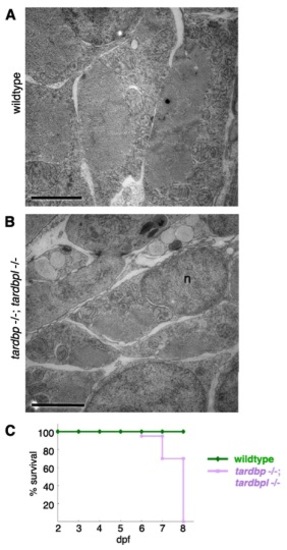
Ultrastructural morphology of myocytes in tardbp-/-;tardbpl-/- mutants analyzed by EM and early lethality. (A) Skeletal muscles of a wild-type embryo display an ordered array of myofibrils surrounded by a highly ordered network of sarcoplasmic reticulum (here shown in cross-sections). Each of the myofibrils is clearly separated by a string of sarcoplasmic reticulum. (B) Skeletal muscle of a tardbp-/-;tardbpl-/- mutant embryo shows a highly disorganized pattern of thinner myofibrils with disorganized network of sarcoplasmic reticulum at 2 dpf.. (Scale bars, 2 μm.) n = nucleus. (C) Kaplan?Meier plot of wild-type (green) and tardbp-/-;tardbpl-/- mutant (purple) embryos. All tardbp-/-;tardbpl-/- mutant embryos analyzed were dead by 8 dpf (n = 20), whereas all wildtype embryos were alive (n = 20). |

