|
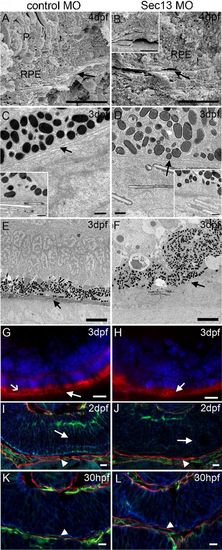
Basal lamina/Bruch′s membrane in Sec13 knock-down embryos.
SEM of internal surfaces of control (A) and Sec13 MO (B, inset for second example) indicating Bruch′s membrane (arrows). P: photoreceptor layer; RPE: retinal pigment epithelium. Scale bars: 10μm. TEM of control (C) and Sec13 MO (D). Arrows: basal lamina. Insets: second examples. Scale bars: 500nm. Tannic acid/uranyl acetate stain of control (E) and Sec13 MO (F). Representative for 2 embryos each. Arrows: collagen-rich layer. Scale bars: 5μm. Immunohistochemical staining for collagen IV of control (G) and Sec13 MO (H) albino fish. Arrows: collagen IV layer; open arrow: auto-fluorescence of outer segments. Scale bars: 10μm. Immunohistochemical staining for laminin (green), actin (red, arrowheads) and 7beta;-catenin (blue) of control (I,K) and Sec13 MO (J,L). Arrows: disorganisation of retinal neuroepithelium shown by laminin and β-catenin labelling. Representative image of 12 embryos. Scale bars: 10μm.
|

