FIGURE SUMMARY
Title

Early stages of retinal development depend on Sec13 function

Authors
Schmidt, K., Cavodeassi, F., Feng, Y., and Stephens, D.J.
Source
Full text @ Biol. Open

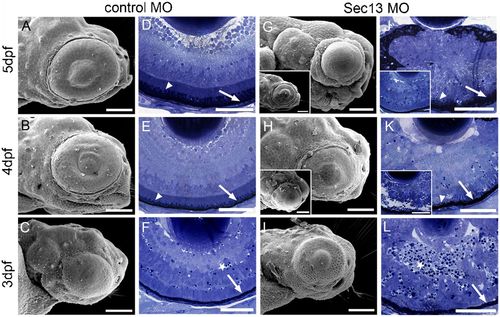
Sec13 morphant phenotype.
SEM of control (A–C) and Sec13 MO littermates (G–I). Scale bars: 100μm. Toluidine Blue-stained 1μm transversal sections of control (D–F) and Sec13 MO (J–L) from a comparable medial level. Scale bars: 50μm. Insets in G,J,H,K show the other, less representative end of the phenotypic spectrum for 4 and 5dpf, respectively. Arrows: RPE; arrowheads: outer segments of photoreceptors; stars: pyknotic nuclei/cell debris.

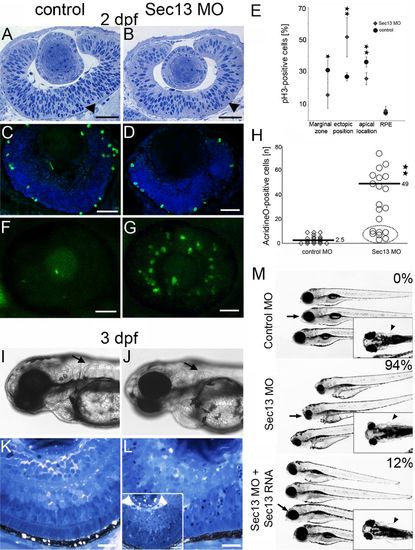
Rescue of Sec13 knock-down phenotype.
Toluidine Blue staining of 1μm transversal sections of control (A) and Sec13 MO (B). pH3 immunostaining on 10μm sections of control (C) and Sec13 MO (D). Quantification of pH3-positive cells (E). 582±113 pH3-positive nuclei were obtained from control morphant eyes versus 174±40 cells in G2/M-phase in Sec13-depleted eyes. AO staining of control (F) and Sec13MO (G). Dotted oval: injected embryos without phenotype therefore excluded from mean values (H). Co-injection of p53 MO with control (I,K) or Sec13 MO (J,L). Live images, representative of 40 embryos each (I,J), and Toluidine Blue stained resin sections (J,K). Rescue experiment (M). Embryonic phenotype was scored according to eye (arrows) and fin phenotype (arrowheads, insets). Number of phenotypic embryos is given as percentage (M, top right). Quantification from at least 60 embryos each from 3 independent experiments. Scale bars: 20μm.

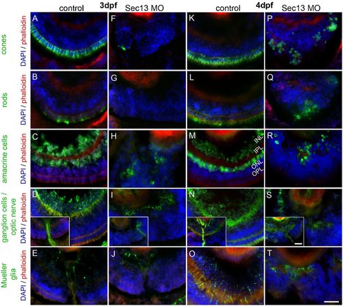
Cellular identities in embryonic Sec13 knock-down eyes (A-T)
Immunohistochemistry of medial transversal sections. The following antibodies were used to identify cell types: zpr-1 (cones), zpr-3 (rods), HuC/D (amacrine and ganglion cells), zn-5 (ganglion cells incl. axons) and zrf-1 (Mueller glia). Insets in D,I,N,S: optic nerve/axons of ganglion cells. INL: inner nuclear layer; IPL: inner plexiform layer; ONL: outer nuclear layer; OPL: outer plexiform layer. Scale bars: 20μm.

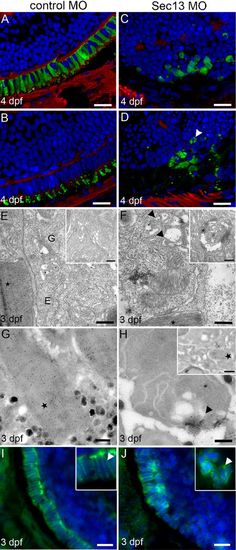
Photoreceptor development and opsin transport in Sec13 morphant eyes.
3D opacity view of control (A,B) and Sec13 MO (C,D) stained for cones (zpr-1, A,C) and opsin (B,D) (green), phalloidin (red) and DAPI (blue). Arrowhead: dotted opsin localisation in Sec13 MO. Scale bars: 10μm. TEM of control (E) and Sec13 MO (F), see insets for additional examples. Stars: outer segments; E: ER; G: Golgi; arrowheads: dilated ER and Golgi. Scale bars: 500nm. Immunogold-labelling of opsin. Control (G) and Sec13MO (H). Arrowhead: accumulation of opsin in cell periphery. Stars: opsin label in outer segments. Scale bars: 500nm. Immunolocalisation of syntaxin-3 in control (I) and Sec13 MO (J). Arrowheads: syntaxin-3 label at plasma membrane. Scale bars: 10μm.

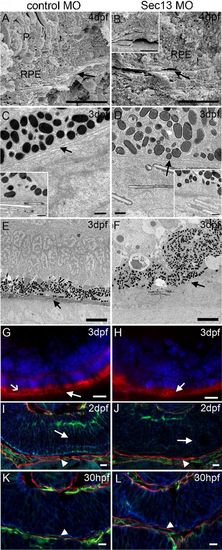
Basal lamina/Bruch′s membrane in Sec13 knock-down embryos.
SEM of internal surfaces of control (A) and Sec13 MO (B, inset for second example) indicating Bruch′s membrane (arrows). P: photoreceptor layer; RPE: retinal pigment epithelium. Scale bars: 10μm. TEM of control (C) and Sec13 MO (D). Arrows: basal lamina. Insets: second examples. Scale bars: 500nm. Tannic acid/uranyl acetate stain of control (E) and Sec13 MO (F). Representative for 2 embryos each. Arrows: collagen-rich layer. Scale bars: 5μm. Immunohistochemical staining for collagen IV of control (G) and Sec13 MO (H) albino fish. Arrows: collagen IV layer; open arrow: auto-fluorescence of outer segments. Scale bars: 10μm. Immunohistochemical staining for laminin (green), actin (red, arrowheads) and 7beta;-catenin (blue) of control (I,K) and Sec13 MO (J,L). Arrows: disorganisation of retinal neuroepithelium shown by laminin and β-catenin labelling. Representative image of 12 embryos. Scale bars: 10μm.

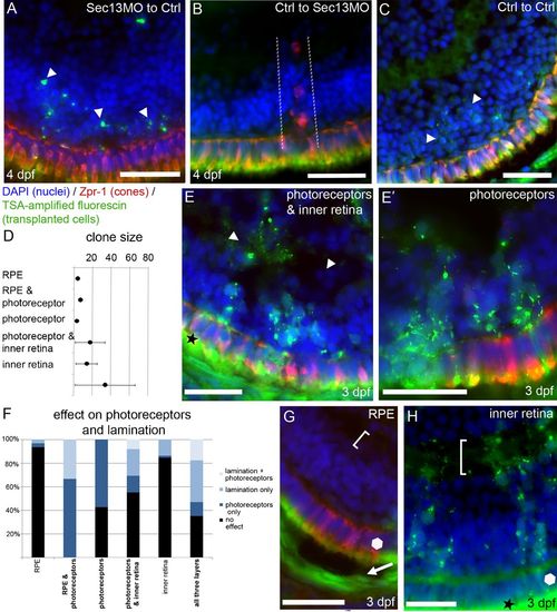
Cell autonomous effect of Sec13 depletion.
Dextran-fluorescin-labelled donor blastomeres were transplanted into host blastulas as indicated. Dextran-fluorescin was detected by TSA amplification (green), cones were identified by zpr-1 labelling (red) and nuclei were stained with DAPI (blue). Examples of blastomere transplants from/to LonAB embryos at 4dpf (A–C). Arrowheads (A): apoptotic morphant cells. Dotted lines (B): photoreceptor cells derived from the Sec13 MO host. Arrowheads (C): transplanted control cells. Blastomere transplantation into albino wild-type embryos (D–H). Sec13 morphant transplants were fluorescin-positive (green) or identified by presence of melanosomes (black cell layer, arrow in G). Graphical depiction (D) of the average number of transplanted blastomeres in each clone (black dots). Phenotypic transplants scored for impact on photoreceptors and/or lamination or no effect shown as fractions of 100% with respect to the number of clones in each category (F). Layers including photoreceptors (bold font) showed more effect. Photoreceptors and lamination: light blue, lamination: medium blue, photoreceptors: dark blue, no effect: black. Examples of transplants (green and/or pigmented) as indicated (E,E2,G,H). Photoreceptors (Zpr-1) in red and nuclei in blue. Arrow: Sec13 derived RPE cells; arrowheads: defects in retinal lamination; brackets: no effect on lamination; polygons: no effect on photoreceptors; star: autofluorescence of outer segments. Scale bars: 20μm.

Acknowledgments
This image is the copyrighted work of the attributed author or publisher, and ZFIN has permission only to display this image to its users. Additional permissions should be obtained from the applicable author or publisher of the image. Full text @ Biol. Open