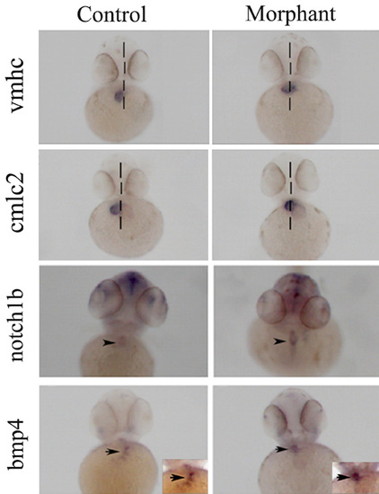
Fig. 5
- ID
- ZDB-FIG-130307-9
- Publication
- Moriarty et al., 2012 - Loss of plakophilin 2 disrupts heart development in zebrafish
- Other Figures
- All Figure Page
- Back to All Figure Page
|
Expression of cardiac morphology marker genes in plakophilin 2 morphant and control morpholino injected embryos. Ventral views of 48 hpf wholemount embryos with (A-D) the midline denoted by a broken line. (A and B) vmhc delineates ventricle specification at 48 hpf in (A) control and (B) morphant embryos. In morphants the ventricle remained at the midline. (C,D) cmlc2 was expressed in both atrium and ventricle at 48 hpf in (C) control and (D) morphant embryos with midline placement in the latter. (E,G) Expression of both notch1b and bmp4 was restricted to the atrio-ventricular boundary (black arrowheads) in control morpholino injected embryo hearts; (F and H) in morphant embryos, both cardiac valve markers were expanded towards the ventricle (black arrows). The numbers of embryos with the displayed phenotype were (A) 67/70 (B) 34/61 (C) 68/70 (D) 28/45 (E) 47/47 (F) 28/49 (G) 49/49 (H) 43/67, numbers of morphant embryos showing the phenotype reflect the proportion of affected embryos (Fig. 1). |
| Fish: | |
|---|---|
| Knockdown Reagent: | |
| Observed In: | |
| Stage: | Long-pec |

