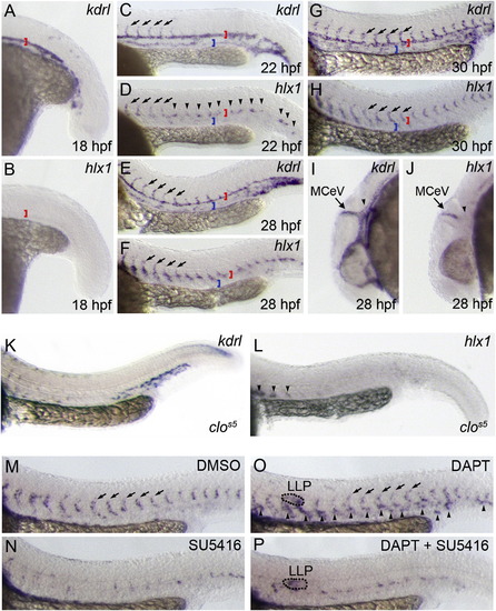
hlx1 Expression Marks Sprouting ECsWhole-mount in situ hybridization analysis of kdrl (A, C, E, G, I, and K) or hlx1 (B, D, F, H, J, and L?P) expression at 18 (A and B), 22 (C and D), 28 (E, F, I, and J), or 30 (G, H, and K?P) hpf in untreated WT embryos (A-J), clos5 mutant embryos (K and L), or WT embryos incubated with DMSO (M), 0.625 μM SU5416 (N), 100 μM DAPT (O), or both 100 μM DAPT and 0.625 μM SU5416 (P) from 22 hpf (A-J; red brackets in A-H mark the DA, whereas blue brackets in C?H mark the cardinal vein). hlx1 is not expressed during early vasculogenic assembly of the DA (B) but is initially expressed in the first-sprouting ISVs (arrows in C and D) and at regions of future angiogenic remodeling (arrowheads in D). At later stages hlx1 expression is almost exclusively restricted to sprouting angiogenic ECs of the ISVs (arrows in E?H) and MCeVs (arrows in I and J) but is excluded from the adjacent nonangiogenic parental tissues of the DA (red brackets in A-H) and PHBC (arrowheads in I and J). (K and L) Both kdrl and hlx1 expressions are reduced or lost in EC-deficient clos5 mutants. Arrowheads in (L) indicate nonendothelial staining of the ventral somite. (M?P) SU5416-mediated suppression of EC sprouting abolished hlx1 expression (N), whereas DAPT promoted ectopic hlx1 expression in nonangiogenic tissues (arrowheads in O). DAPT-induced expression of hlx1 was lost upon coincubation of embryos with the VEGFR inhibitor, SU5416 (P) (LLP, putative lateral line primordium).
|

