FIGURE

Fig. S3

ID
ZDB-FIG-121212-1
Publication
Herbert et al., 2012 - Determination of Endothelial Stalk versus Tip Cell Potential during Angiogenesis by H2.0-like Homeobox-1
Other Figures
All Figure Page
Back to All Figure Page
Fig. S3

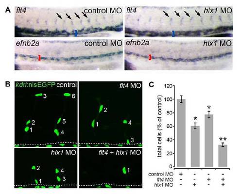
Flt4-Dependent TC Formation Appears Unaffected in the Absence of Hlx1, Related to Figures 3 and 4
(A) Whole-mount in situ hybridization analysis of flt4 and efnb2a expression at 30 hpf in control and hlx1 MO-injected embryos. Hlx1 knockdown did not detectibly disrupt flt4 expression in sprouting TCs (arrows) and the CV (blue brackets) or efnb2a expression in the DA (red brackets).
(B-C) Lateral views of Tg(kdrl:nlsEGFP)zf109 (B; dotted line represents position of DA) embryos and quantification of ISV EC numbers (C) at 30 hpf upon injection of embryos with the indicated combinations of control MO, flt4 MO and hlx1 MOs. Upon Hlx1 knockdown EC sprouting remains highly Flt4-dependent. (n = at least 13 embryos). Error bars represent mean ± SEM. (*P < 0.05 versus control MO; **P < 0.05 versus flt4 MO or hlx1 MO).

Expression Data

Expression Detail
Antibody Labeling
Phenotype Data

Phenotype Detail
Acknowledgments
This image is the copyrighted work of the attributed author or publisher, and ZFIN has permission only to display this image to its users. Additional permissions should be obtained from the applicable author or publisher of the image. Full text @ Curr. Biol.