Fig. S6
- ID
- ZDB-FIG-121031-45
- Publication
- Shimizu et al., 2012 - Visualization and exploration of Tcf/Lef function using a highly responsive Wnt/beta-catenin signaling-reporter transgenic zebrafish
- Other Figures
- All Figure Page
- Back to All Figure Page
|
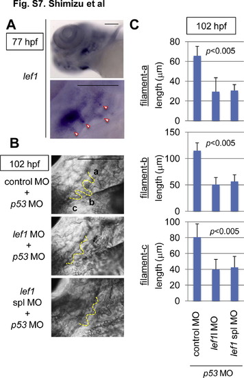
Lef1 was required for gill filament growth. (A) Whole-mount in situ hybridization staining for lef1 in 77-hpf zebrafish embryos. Panels show lower left-side head views (upper panel) and magnified gill area views (lower panel) of embryos, with the anterior side to the left. The gill filament bud-expressing lef1 mRNA is indicated with arrowheads. Scale bar, 100 μm. (B, C) lef1 knockdown reduced the gill filament length. Zebrafish embryos were injected with control MO (n = 9), lef1 MO (n = 13), or lef1 spl MO (n = 9) with p53 MO, as indicated. (B) Panels show gill area views of 120-hpf MO-injected embryos, with the anterior side to the left. The outlines of gill filaments and their buds are shown with yellow broken lines. Scale bar, 100 μm. Three gill filaments budded from branchial arch 1 are labeled a, b, and c, and their lengths are shown. (C) Graphs show the lengths of the labeled gill filaments, as indicated in B, of 102-hpf MO-injected embryos (n = 11 each). Data are mean ± SD. The p value was calculated by Student′s t test. |
Reprinted from Developmental Biology, 370(1), Shimizu, N., Kawakami, K., and Ishitani, T., Visualization and exploration of Tcf/Lef function using a highly responsive Wnt/beta-catenin signaling-reporter transgenic zebrafish, 71-85, Copyright (2012) with permission from Elsevier. Full text @ Dev. Biol.

