|
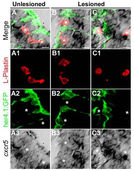
cxcr5 is not expressed in L-Plastin-positive cells in the adult zebrafish telencephalon. (A) cxcr5 in situ hybridization coupled to immunohistochemistry for L-Plastin (red, marking macrophages and microglia) and her4.1:green fluorescent protein (GFP) (green, marking the radial glial cells) in the unlesioned telencephalons. White asterisks indicate the L-Plastin cells. (A1-A3) Individual channels for L-Plastin, her4.1:GFP and cxcr5, respectively. (B) cxcr5 in situ hybridization coupled to immunohistochemistry for L-Plastin and her4.1:GFP in the lesioned telencephalons (dorsolateral region). White asterisks indicate the L-Plastin cells. (B1-B3) Individual channels for L-Plastin, her4.1:GFP and cxcr5, respectively. (C) cxcr5 in situ hybridization coupled to immunohistochemistry for L-Plastin and her4.1:GFP in the lesioned telencephalons (dorsomedial region). White asterisks indicate the L-Plastin cells. (C1-C3) Individual channels for L-Plastin, her4.1:GFP and cxcr5, respectively.
|