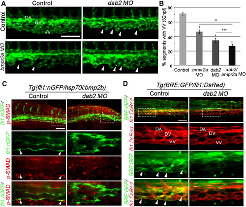
Dab2 Functionally Interacts with Bmp2b Signal to Promote Caudal Vein Plexus Formation (A) Fluorescent images of 32 hpf control, dab2, bmpr2a, and dab2/bmpr2a MO-injected embryos. Arrowheads point to the regions that fail to connect to neighboring sprouts. Scale bar is 100 μm. See also Figure S2A.(B) Quantification of ventral vein (VV) defects. n = 3, and total number of embryos was 54 (control), 32 (bmpr2a MO), 26 (dab2 MO), or 35 (dab2/bmpr2a MO). p < 0.001 in all cases, except p value against dab2 MO-injected embryos, where p = 0.05. comparison with control MO-injected embryos; comparison with bmpr2a MO-injected embryos; and comparison with dab2 MO-injected embryos. Error bars represent SEM.(C) Confocal micrographs of control or dab2 MO-injected 45 hpf Tg(hsp70l:bmp2b) embryos, showing p-Smad-1,5/8 (red) and endothelial cells (green). Arrowheads point to p-Smad-1,5/8 within venous endothelial cells. Scale bar is 100 μm.(D) Confocal micrographs of control or dab2 MO-injected 45 hpf Tg(BRE:GFP);Tg(fli1:DsRed) embryos. Arrowheads point to GFP expression by BRE. Scale bar is 100 μm. DA, dorsal aorta; DV, dorsal vein; VV, ventral vein.
|
