
Fig. S5
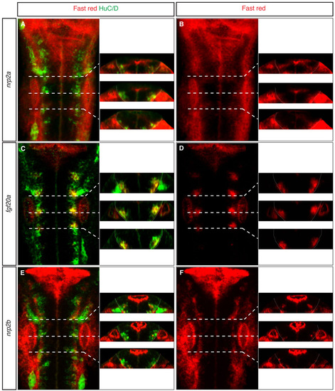
fgf20a and nrp2a expression colocalise. Expression pattern of (A,B) nrp2a, (C,D) fgf20a and (E,F) nrp2b. Left side of each panel shows confocal images of dorsal views of 24-hpf zebrafish hindbrain, anterior to the top. Right side shows z-reconstruction at AP positions marked by white dashed lines of the same embryo. White line delimits the neural tube position. The fluorescent in situ hybridisation signal was generated by Fast Red staining (red) and combined with Hu antibody staining (green). This approach was taken because the signals were too weak to achieve double in situ hybridisation staining. Orientation of embryos as Fig. 1. |