Fig. 1
|
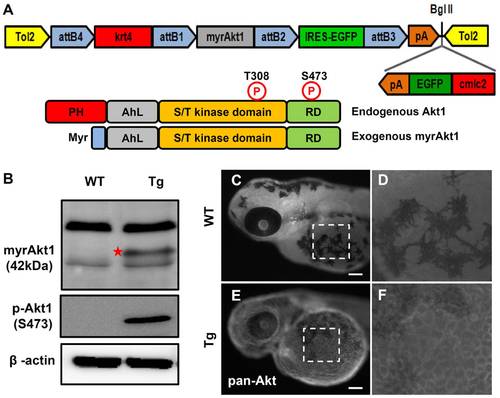
Generation of Tg(krt4:Hsa.myrAkt1)cy18. (A) The schematic diagram in the upper panel shows the configuration of the pDestTol2CG2-krt4-myrAkt1-IRES-EGFP-pA plasmid used to generate Tg(krt4:Hsa.myrAkt1)cy18. The parental vector of pDestTol2CG2, which contains cmlc2-EGFP-pA mini-gene, helps transgenic progeny to express green fluorescent protein in the heart. The lower panel illustrates the domain structure of the wild-type and constitutively active form of human Akt1. (B) Western blot analysis of protein lysate extracted from adult tail fins shows that the exogenous human myrAkt1 (indicated by star) is only detectable in Tg(krt4:Hsa.myrAkt1)cy18. β-actin served as a loading control. Whole-mount immunostaining of wild-type (C) or Tg(krt4:Hsa.myrAkt1)cy18(E) embryos aged 3 days post-fertilization using pan-Akt antibody. The yolk sac region highlighted by a dotted line in C and E is magnified in E and F, respectively. PH, Pleckstrin homology; RD, regulation domain; Myr, myristylation signal; WT, wild type; Tg, Tg(krt4:Hsa.myrAkt1)cy18. Scale bar = 100 μm. |
| Antibodies: | |
|---|---|
| Fish: | |
| Anatomical Terms: | |
| Stage Range: | Protruding-mouth to Adult |

