Fig. S5
- ID
- ZDB-FIG-120601-24
- Publication
- Chablais et al., 2012 - The regenerative capacity of the zebrafish heart is dependent on TGFβ signaling
- Other Figures
- All Figure Page
- Back to All Figure Page
|
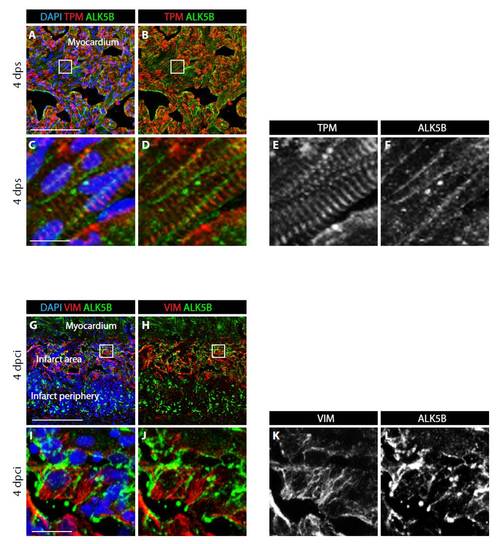
Alk5b is expressed in cardiomyocytes and vimentin-positive cells of cryoinjured hearts. (A-D) High magnification of the ventricle after sham surgery immunostained for Tropomyosin (red), Alk5b (green) and DAPI (blue). (C,D) Higher magnifications of the framed area in the corresponding upper panels. (E,F) Single grayscale channel showing Tropomyosin (E) and Alk5b (F). In uninjured myocardium, linear pattern of Alk5b staining corresponds to the plasma membrane of cardiomyocytes. Tropomyosin staining labels striated muscle fibers. (G-J) High-magnification images of a post-infarct zone from cryoinjured heart at 4 dpci, stained for the fibroblast marker Vimentin (red), Alk5b (green) and DAPI (blue). (I,J) Higher magnifications of the framed area in the corresponding upper panels. (K,L) Single grayscale channel showing Vimentin (K) and Alk5b (L). Vimentin-positive cells express transmembrane receptor Alk5b. Scale bars: 100 μm in A,G; 10 μm in C,I. |

