
Fig. S1
- ID
- ZDB-FIG-120601-20
- Publication
- Chablais et al., 2012 - The regenerative capacity of the zebrafish heart is dependent on TGF? signaling
- Other Figures
- All Figure Page
- Back to All Figure Page
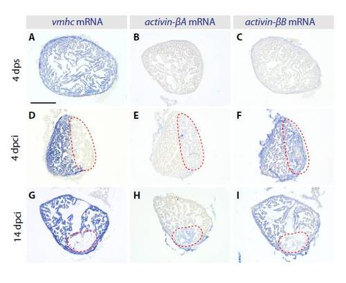
Expression of Activin-β ligands in cryoinjured heart. In situ hybridization of consecutive heart sections with vmhc, activin-βA and activin-βB mRNA antisense probes (blue staining) at different phases of heart regeneration. (A-C) In uninjured sham operated hearts at 4 dps, vmhc (A) is expressed in the entire ventricle, whereas activin-βA (B) and activin-βB (C) do not display significant expression. (D-F) In regenerating hearts at 4 dpci, the absence of vmhc (D) marks the injury area (red dashed line). activin-βA (E) is detected in the infarct zone. activin-βB (F) is induced in the whole infarcted ventricle including the myocardium and the injury site. (G-I) At 14 dpci, the post-infarct area (red dashed lines) is smaller than at 4 dpci. activin-βA (H) is detected in the injury area. activin-βB (I) is expressed in the whole infracted ventricle. Scale bar: 300 μm. |